
Maintaining strong bones allows you to stay active, independent, and confident throughout your retirement years. A simple bone density scan provides an accurate window into your skeletal strength, helping you detect weakening bones long before a painful fracture occurs. Over 54 million older Americans live with low bone mass or osteoporosis, yet many remain unaware of their condition until a simple slip causes serious injury. Recognizing early physical clues and personal risk factors empowers you to take proactive control of your health. Reviewing the core indicators for testing gives you the knowledge needed to discuss your bone health with your physician today.
Understanding the Basics of Bone Density Scans for Seniors
Your bones are living, dynamic tissues that continuously break down and rebuild throughout your life. During your youth, bone formation outpaces bone loss; however, as you age, this natural cycle shifts. After menopause in women and during later decades in men, bone breakdown begins to outpace regeneration. Over time, this imbalance leads to porous, fragile bones—a progressive condition known as osteoporosis.
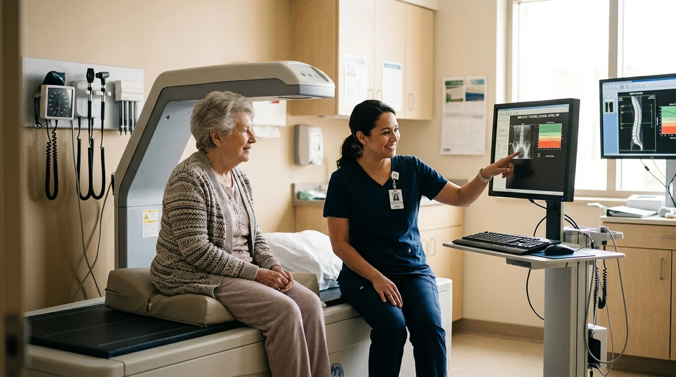
A dual-energy X-ray absorptiometry scan, commonly referred to as a DEXA scan, represents the gold standard for measuring your bone mineral density. This non-invasive, completely painless imaging test uses very low levels of radiation to scan your hip and lumbar spine. In fact, a single DEXA scan exposes you to less radiation than a standard chest X-ray or an airplane flight across the country. The scan takes approximately ten to fifteen minutes while you lie comfortably on a padded table.
Doctors evaluate your scan results using a standardized measurement called a T-score, established by the World Health Organization. Your T-score compares your current bone density to the optimal peak bone mass of a healthy young adult:
Normal Bone Density: A T-score of -1.0 or higher indicates healthy, robust bone mineral levels.
Low Bone Mass (Osteopenia): A T-score between -1.0 and -2.5 reveals thinning bones that require protective lifestyle measures and medical monitoring.
Osteoporosis: A T-score of -2.5 or lower confirms significant bone loss, which places you at a higher risk for unexpected fractures.
Following established osteoporosis scan guidelines ensures that you catch bone thinning early. Statistics show that approximately one in two women and up to one in four men over age 50 will suffer an osteoporosis-related fracture. Recognizing the physical markers and clinical milestones helps you protect your mobility for decades to come.

Practical Tips for a Healthier, Happier Life
Knowing when to ask for a DEXA scan empowers you to make proactive choices rather than reacting to a sudden injury. Review these seven clear signs to determine if you should request a bone density evaluation during your next medical checkup.
1. You Have Reached Key Milestone Screening Ages
Age remains the single most influential factor in skeletal remodeling. Official bone density testing recommendations from leading medical organizations suggest universal screening thresholds for older adults. The U.S. Preventive Services Task Force and the Bone Health and Osteoporosis Foundation recommend routine DEXA screenings for all women aged 65 and older. For men, the Bone Health and Osteoporosis Foundation and the Endocrine Society advise routine testing starting at age 70.
Postmenopausal women between the ages of 50 and 64 should also evaluate their bone density scan eligibility if they have one or more clinical risk factors, such as a history of smoking, a slender frame, or a family history of fractures. Reaching these milestone ages is a clear reason to request a baseline scan, even if you feel completely healthy.
2. You Notice Unexplained Height Loss or Postural Changes
A common physical indicator of bone thinning is a noticeable decrease in height. If you have lost 1.5 inches or more from your peak adult height—or half an inch within a single calendar year—you may be experiencing silent vertebral compression fractures. These tiny micro-fractures in the spine often develop without causing sharp pain; instead, they slowly compress your spinal column.
In addition to height reduction, a rounding of your upper back and shoulders, sometimes referred to as kyphosis, serves as one of the physical signs you need a bone scan. When spinal vertebrae weaken, the upper spine curves forward under normal daily pressure. If your clothes suddenly fit differently around your torso, or your posture stoops forward despite your best efforts to stand straight, schedule a bone density evaluation.
3. You Experienced a Broken Bone from a Minor Fall
Healthy adult bones can absorb everyday bumps and minor slips. If you trip over a rug, step off a curb awkwardly, or slip on a flat surface and break a bone, doctors classify this as a fragility fracture. Breaking a wrist, hip, pelvis, or upper arm from a fall at standing height or less after age 50 is a significant clinical signal that your bone architecture has weakened.
A low-impact fracture warrants an immediate DEXA scan regardless of your age or previous health history. Suffering one fragility fracture doubles your risk of experiencing a second break within the following year; therefore, a prompt scan allows your physician to implement therapeutic interventions that protect your remaining bone mass.
4. You Take Medications Known to Weaken Bone Structure
Several routine prescription medications accelerate bone breakdown when used over long periods. Corticosteroids, such as oral prednisone, represent the most common culprits. Taking five milligrams or more of prednisone daily for three consecutive months directly impairs your body’s bone-building cells and reduces intestinal calcium absorption.
Other medications that contribute to progressive bone loss include long-term proton pump inhibitors used for acid reflux, certain anti-seizure drugs, aromatase inhibitors for breast cancer, and androgen deprivation therapy for prostate cancer. If you rely on any of these medications for chronic symptom management, ask your healthcare provider about scheduling a baseline DEXA scan.
5. You Experienced Early Menopause or Hormonal Deficiencies
Estrogen plays a vital role in slowing bone breakdown and preserving mineral balance. When estrogen production drops dramatically—whether through natural menopause before age 45 or the surgical removal of both ovaries—bone resorption accelerates rapidly. Women can lose up to twenty percent of their total bone mass during the five to seven years immediately following menopause.
Hormone levels influence male bone health as well. Men who experience low testosterone levels, whether due to aging, hormonal disorders, or medical treatments, face an elevated risk of bone thinning. If you underwent early menopause or manage hormonal imbalances, discuss a preventative DEXA scan with your doctor.
6. You Have a Naturally Slender Build or Low Body Weight
Having a petite frame or low body mass index is an established independent risk factor for accelerated bone thinning. If you weigh under 127 pounds or have a body mass index below 19, your body naturally possesses less peak bone mass to draw upon as you grow older. Smaller bones naturally have less bone tissue overall.
Furthermore, individuals with low body fat produce lower levels of estrogen, which fat cells naturally synthesize in modest amounts after menopause. If you have maintained a very light frame throughout your adult life, your physician will likely recommend earlier bone density testing to establish your baseline values.
7. You Have a Strong Family History of Osteoporosis or Fractures
Genetics account for sixty to eighty percent of your overall peak bone mass potential. If one of your parents suffered a broken hip after a minor slip, lost substantial height in their older years, or received an osteoporosis diagnosis, your personal risk increases significantly.
Family medical history offers valuable insights into your inherited bone density potential and your risk for future fractures. Sharing this background with your doctor helps them assess whether you should undergo bone density testing earlier than standard age guidelines recommend.
Daily Habits to Protect and Nourish Your Bones
While a bone density scan provides critical diagnostic insight, daily holistic habits actively strengthen your skeletal structure. Pairing clinical awareness with proactive lifestyle choices creates a solid foundation for healthy aging.
Nourish with Bone-Building Nutrients: Aim for 1,200 milligrams of daily calcium through whole foods such as plain yogurt, kefir, sardines with bones, collard greens, almonds, and fortified plant milks. Pair your calcium intake with 800 to 1,000 international units of Vitamin D3 daily to support efficient mineral absorption in your digestive tract.
Engage in Weight-Bearing Movement: Bones respond to physical demand by growing denser. Incorporate thirty minutes of weight-bearing exercise—such as brisk walking, climbing stairs, dancing, or light resistance band training—into your routine five days per week.
Practice Balance and Stability: Protecting your bones also means avoiding falls. Gentle balance exercises, including Tai Chi, heel-to-toe walking, and single-leg balance drills near a sturdy counter, enhance your neuromuscular coordination and stability.

Important Safety Precautions to Keep in Mind
Maintaining bone health requires a mindful, cautious approach to your home environment and exercise routine. If you suspect your bones have weakened, or if you have received an osteopenia diagnosis, take deliberate steps to minimize physical strain and reduce fall hazards.
Evaluate your living space for everyday tripping risks. Secure loose throw rugs with non-skid backing or remove them entirely. Ensure all staircases, hallways, and bathrooms have bright lighting, and install sturdy grab bars inside showers and beside toilets. Always wear supportive, closed-toe shoes with non-slip soles indoors rather than walking in smooth socks or loose slippers.
Modify your physical exercise routines to protect your spine. If you have low bone density, avoid high-impact jumping exercises and activities that require deep forward spinal bending, such as standard sit-ups, toe touches, or heavy abdominal crunches. Avoid forceful spinal twisting movements, which can place excessive pressure on delicate vertebrae; instead, focus on exercises that promote an elongated, upright posture.
Be thoughtful regarding dietary supplements. While adequate calcium is vital, consuming excessive calcium via high-dose pills can increase the risk of kidney stones and arterial calcification. Focus primarily on obtaining calcium through nutrient-dense foods, and use modest supplements only to bridge any remaining dietary gaps under your doctor’s supervision.

When to Speak with a Healthcare Professional
A DEXA scan is a straightforward, accessible preventive tool that gives you clear facts about your skeletal health. Medicare Part B covers a central DEXA bone density scan once every 24 months at zero out-of-pocket cost for qualified beneficiaries, including estrogen-deficient individuals, people taking long-term corticosteroids, and those with known spinal abnormalities. For individuals paying out of pocket, the national average self-pay cost at outpatient imaging centers typically ranges between $100 and $250.
Schedule an appointment with your healthcare provider promptly if you notice sudden, severe back pain, experience unexplained postural changes, or have recently suffered any broken bone from a minor fall. When you visit your doctor, bring a complete list of your current medications, any history of fractures, and details regarding your family medical background. Ask your doctor whether a central DEXA scan is appropriate for your age and personal health profile.
For authoritative, trustworthy health information, always consult official sources like the National Institute on Aging (NIA) and MedlinePlus from the National Library of Medicine. The CDC also provides excellent resources for older adults.
Frequently Asked Questions
Does a bone density scan hurt, and how long does it take?
A DEXA bone density scan is completely painless and non-invasive. You do not need injections, medications, or uncomfortable physical restraints. You simply lie flat on a cushioned examination table while an open mechanical arm passes slowly over your lower spine and hips. The entire scanning process usually takes between ten and fifteen minutes, and you can immediately resume your normal daily activities.
What is the difference between a peripheral bone scan and a central DEXA scan?
Peripheral scans, sometimes offered at community health fairs or pharmacies, measure bone density in peripheral areas like your finger, wrist, or heel. While these portable screenings provide a helpful general indicator, they cannot officially diagnose osteoporosis or accurately track medication response. A central DEXA scan measures the hip and lumbar spine, providing the precise clinical data required for medical treatment decisions.
Can you rebuild bone density naturally after age 60?
While you cannot completely reverse severe osteoporosis through lifestyle adjustments alone, you can significantly slow bone breakdown, stabilize your bone mineral density, and stimulate bone maintenance. Combining adequate daily calcium and Vitamin D with consistent resistance training and weight-bearing movement helps preserve bone architecture and substantially lowers your risk of falling and fracturing a bone.
How often should seniors repeat a bone density scan?
Most healthcare providers recommend repeating a DEXA scan every two years for individuals diagnosed with osteopenia or osteoporosis, or for those taking prescription bone-strengthening medications. This 24-month interval aligns with standard Medicare coverage rules and gives your body sufficient time to demonstrate measurable skeletal changes in response to lifestyle improvements or therapies.
Disclaimer: This article is for informational purposes only and does not constitute medical advice. Always seek the advice of your physician or other qualified health provider with any questions you may have regarding a medical condition.